
SELECTED PUBLICATIONS

LOXHD1 is indispensable for maintaining TMC1 auditory mechanosensitive channels at the site of force transmission.
Wang P, Miller KK, He E, Dhawan SS, Cunningham CL, Grillet N.
Nat Commun. 2024 Sep 10;15(1):7865.
PMID: 39256406 Journal Featured
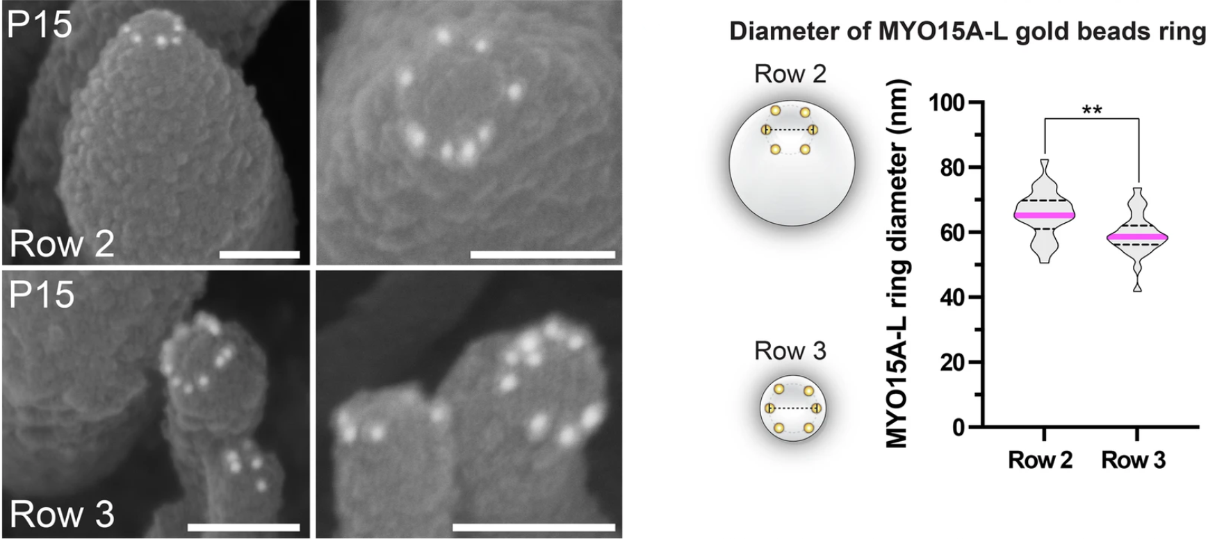
SUB-immunogold-SEM reveals nanoscale distribution of submembranous epitopes.
Miller KK, Wang P, Grillet N.
Nat Commun. 2024 Sep 10;15(1):7864.

Single-cell transcriptomic atlas reveals increased regeneration in diseased human inner ear balance organs.
Wang T, Ling AH, Billings SE, Hosseini DK, Vaisbuch Y, Kim GS, Atkinson PJ, Sayyid ZN, Aaron KA, Wagh D, Pham N, Scheibinger M, Zhou R, Ishiyama A, Moore LS, Maria PS, Blevins NH, Jackler RK, Alyono JC, Kveton J, Navaratnam D, Heller S, Lopez IA, Grillet N., Jan TA, Cheng AG.
Nat Commun. 2024 Jun 6;15(1):4833.
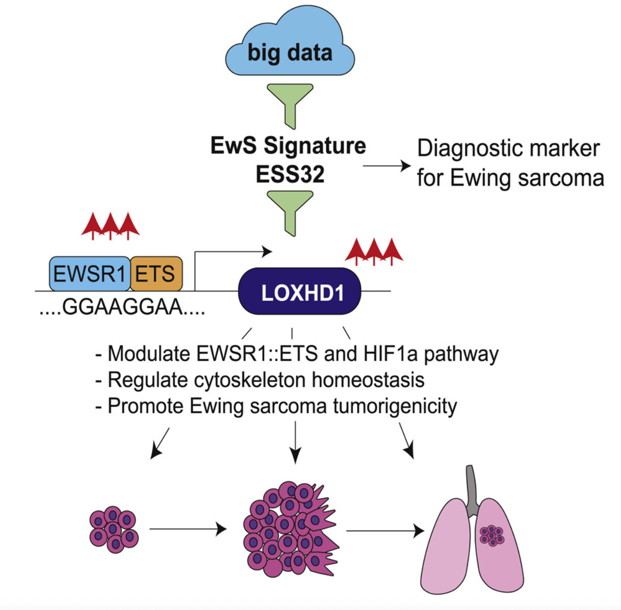
Oncofusion-driven de novo enhancer assembly promotes malignancy in Ewing sarcoma via aberrant expression of the stereociliary protein LOXHD1.
Deng Q, Natesan R, Cidre-Aranaz F, Arif S, Liu Y, Rasool RU, Wang P, Mitchell-Velasquez E, Das CK, Vinca E, Cramer Z, Grohar PJ, Chou M, Kumar-Sinha C, Weber K, Eisinger-Mathason TSK, Grillet N., Grünewald TGP, Asangani IA.
Cell Rep. 2022 Jun 14;39(11):110971.

Dimensions of a Living Cochlear Hair Bundle.
Miller KK, Atkinson P, Mendoza KR, Ó Maoiléidigh D*, Grillet N*.
Front Cell Dev Biol. 2021;9:742529.

Loxhd1 Mutations Cause Mechanotransduction Defects in Cochlear Hair Cells.
Trouillet A, Miller KK, George SS, Wang P, Ali NE, Ricci A, Grillet N.
J Neurosci. 2021 Apr 14;41(15):3331-3343.

Mechanosensory hair cells express two molecularly distinct mechanotransduction channels.
Wu Z*, Grillet N* Zhao B, Cunningham C, Harkins-Perry S, Coste B, Ranade S, Zebarjadi N, Beurg M, Fettiplace R, Patapoutian A, Mueller U.
Nat Neurosci. 2017 Jan;20(1):24-33.

Neuroplastin Isoform Np55 Is Expressed in the Stereocilia of Outer Hair Cells and Required for Normal Outer Hair Cell Function.
Zeng WZ*, Grillet N*, Dewey JB, Trouillet A, Krey JF, Barr-Gillespie PG, Oghalai JS, Müller U.
J Neurosci. 2016 Aug 31;36(35):9201-16.

TMIE is an essential component of the mechanotransduction machinery of cochlear hair cells.
Zhao B, Wu Z, Grillet N., Yan L, Xiong W, Harkins-Perry S, Müller U.
Neuron. 2014 Dec 3;84(5):954-67.

TMHS is an integral component of the mechanotransduction machinery of cochlear hair cells.
Xiong W, Grillet N, Elledge HM, Wagner TF, Zhao B, Johnson KR, Kazmierczak P, Müller U.
Cell. 2012 Dec 7;151(6):1283-95.
Regulation of PCDH15 function in mechanosensory hair cells by alternative splicing of the cytoplasmic domain.
Webb SW, Grillet N, Andrade LR, Xiong W, Swarthout L, Della Santina CC, Kachar B, Müller U.
Development. 2011 Apr;138(8):1607-17.

Mutations in LOXHD1, an evolutionarily conserved stereociliary protein, disrupt hair cell function in mice and cause progressive hearing loss in humans.
Grillet N*, Schwander M*, Hildebrand MS, Sczaniecka A, Kolatkar A, Velasco J, Webster JA, Kahrizi K, Najmabadi H, Kimberling WJ, Stephan D, Bahlo M, Wiltshire T, Tarantino LM, Kuhn P, Smith RJ, Müller U.
Am J Hum Genet. 2009 Sep;85(3):328-37.

Harmonin mutations cause mechanotransduction defects in cochlear hair cells.
Grillet N*, Xiong W*, Reynolds A*, Kazmierczak P, Sato T, Lillo C, Dumont RA, Hintermann E, Sczaniecka A, Schwander M, Williams D, Kachar B, Gillespie PG, Müller U.
Neuron. 2009 May 14;62(3):375-87.
PROTOCOLS
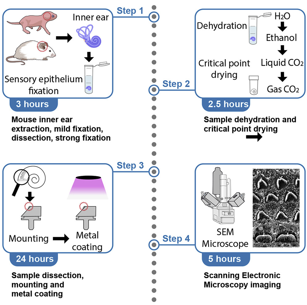
High-resolution imaging of the mouse-hair-cell hair bundle by scanning electron microscopy.
Grillet N.
STAR Protoc. 2022 Mar 18;3(1):101213.
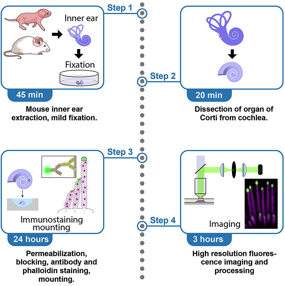
High-resolution immunofluorescence imaging of mouse cochlear hair bundles.
Miller KK, Wang P, Grillet N.
STAR Protoc. 2022 Jun 17;3(2):101431.
REVIEWS
The genetics of progressive hearing loss: a link between hearing impairment and dysfunction of mechanosensory hair cells.
Müller U, Grillet N.
Future Neurol. 2010 Jan 1;5(1):9-12.

The mechanotransduction machinery of hair cells.
Grillet N, Kazmierczak P, Xiong W, Schwander M, Reynolds A, Sakaguchi H, Tokita J, Kachar B, Müller U.
Sci Signal. 2009 Aug 25;2(85):pt5.